Advantages Of Staining Specimens To Be Viewed Under A Microscope
Staining allows different structural components of the cells to be visualized including the cytoplasm cell wall and membranes. Stains or dyes contain salts made up of a positive ion and a negative ion.

Image Result For Diatoms Under Microscope 40x Diatom Peace Symbol Marine
It can be used to view cells in large quantities of the medium than in small specimen quantities on a glass slide under a coverslip.

Advantages of staining specimens to be viewed under a microscope. The most basic reason that cells are stained is to enhance visualization of the cell or certain cellular components under a microscope. Observation When viewed under the microscope students will notice tiny dark spots in the cytoplasm of the organism while the cytoplasm is lightly stained. It makes the microorganisms easily visible under the microscope.
The most basic reason that cells are stained is to enhance visualization of the cell or certain cellular components under a microscope. The steps of the Gram stain procedure are listed. The Gram stain procedure is a differential staining procedure that involves multiple steps.
Dehydrating a sample with alcohol and then placing it in a mould of resin or wax to form a hard block before slicing it with a microtome into thin slices. Staining the specimen One small problem is that most organic specimens such as cells and bacteria are either fully transparent or opaque which results in a drastically low contrast when viewed under the microscope. Common stains used on bacteria include crystal violet methylene blue and safranin.
Its shape size and arrangement can be viewed comfortably. Moreover staining kills the otherwise harmful bacteria which can not be handled due to risk of disease. Liji Thomas MD Reviewed by Georgios Christofidis PhD.
Advantages of Brightfield Microscope It is simple to use with few adjustments involved while viewing the image. When looking at bacteria under the microscope much of the bacteria can appear transparent without staining. The optics of the microscope do not alter the color of the specimen.
Using chemicals like formaldehyde to preserve specimens in a near-natural state. Keep in mind that bacteria which are present in an unstained smear will be invisible when you view it using a light microscope but once stained their arrangement and morphology will make it able to be observed. They give images more contrast and allow cells to be classified according to their shape morphology.
It can be used to view the cell tissues in their original vessel which are larger than microscopic slides which makes it better than the upright microscope which only views specimens in small microscopic slides. Stains or dyes contain salts made up of a positive ion and a negative ion. The other uncolored ion is called the counterion.
It can be used to view both stained and unstained. Thus making use of certain dyes and staining techniques is. In addition to fixation staining is almost always applied to color certain features of a specimen before examining it under a light microscope.
It was developed by Danish microbiologist Hans Christian Gram in 1884 as an effective method to distinguish between bacteria with different types of cell walls and even today it remains one of the most frequently used staining techniques. Microscopy Staining solution Microscopy stains enhance the visualization of cells and cell parts under a light microscope. Direct observation of the organism without staining has a great advantage in that the amoebae are still alive and.
Because of the microscopy requirements options for preparing specimens are limited to. Stains or dyes contain salts made up of a positive ion and a negative ion. Cells may also be stained to highlight metabolic processes or to differentiate between live and dead cells in a sample.
Cells may also be stained to highlight metabolic processes or to differentiate between live and dead cells in a sample. Fixing sectioning staining mounting. These kinds of specimens are difficult to see under a regular microscope.
In addition to fixation staining is almost always applied to color certain features of a specimen before examining it under a light microscope. Whole-mounts where an entire organism or structure is small enough or thin enough to be placed directly onto a microscope slide eg a small unicellular or multicellular organism or a membrane that can be stretched thinly on to a slide. Staining is the most common technique to enhance the visibility of your specimens by increasing the contrast.
Depending on the type of dye the positive or the negative ion may be the chromophore the colored ion. In addition to fixation staining is almost always applied to color certain features of a specimen before examining it under a light microscope. It fixes bacteria on the slide for proper visualisation.
When samples are viewed under the light of a microscope details can be viewed more clearly using light-absorbing stains.

Volvox Microscopic Photography Petri Dish Microscopic Organisms

Special Stains Renal Cell Carcinoma Basement Membrane Stains

Chapter 2 Flashcards Quizlet

What Is The Microscope Field Of View Microscope Field Views

Cellular Respiration Worksheet Answers Printable Worksheets Are A Valuable School Room Tool In 2021 Cellular Respiration Biology Worksheet Photosynthesis Worksheet

Gram Staining Amrita University Microbiology Medical Laboratory Science Student Medical Laboratory Scientist

Wuchereria Bancrofti Medical Laboratory Science Bancroft Medical Laboratory
Advantages And Disadvantages Of Electron Microscopy

Optical Microscopy Specimen Preparation Staining And Quantitative Analysis Conduct Science

Tardigrade In Dark Field Microscope Microscopy Microscopic Photomicrography Science Tardigrade Olympus Canon Wa Microscopy Tardigrade Abstract Artwork

Example Of Phase Contrast Image Contrast Microscope Biology Class
Penicillium Microscopy Requirements Preparation Observation Microscopy Fungi Fungi Art
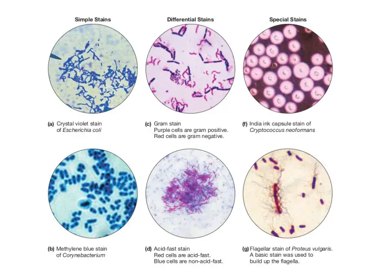
Types Of Staining Techniques Used In Microbiology Microbe Online
Https Oer Galileo Usg Edu Cgi Viewcontent Cgi Filename 0 Article 1016 Context Biology Textbooks Type Additional

Pin On Nature

Print Of Bread Mould Under A Microscope White Mould Fungus Mucor Sp Germany Europe Bread Mold Things Under A Microscope Extreme Close Up

Bio 304 Ecology Evolution Genetics Cell Division Cell Biology Plant Science
Posting Komentar untuk "Advantages Of Staining Specimens To Be Viewed Under A Microscope"